The Division of Emergency Neurosciences (DEN) and Critical Care Research was developed in order to support local and national clinical trials research. This is a ED based consortium composed of physicians from Emergency Medicine, Cardiology, Neurosurgery, Neurology, Diagnostic Imaging, Surgery, and Neurointensive Care. The program sponsors five full time Research Assistants to assist in recruitment and subject retention 24 hours a day, 7 days per week.
The DEN supports active phase I and phase II studies, as well as phase III national clinical trials, such as: POINT, ProTECT III/BIO-ProTECT, iDef, ESETT, ATACH-II, and DEFUSE 3.
In 2017, the DEN has expanded to include research in general critical care and resuscitative medicine. Dr. Lisa H. Merck MD MPH was awarded grant funding as a PI for the SIREN | CORE-EM Hub Alliance, within the new SIREN U24 grant, NIH 1 U24 NS100673-01. SIREN establishes a large, multicenter emergency medicine clinical trial network for the next five years. The Network for Emergency Care Clinical Trials: Strategies to Innovate EmeRgENcy Care Clinical Trials Network (SIREN) to support resuscitative research in cardiopulmonary, neurologic, and traumatic injuries will include 11 hubs nationally.
This represents a tremendous effort of interdisciplinary collaboration within the Brown / Lifespan community, as well as with our national colleagues. The CORE-EM Alliance serves an encatchment area of over 53 million patients across centers from Rhode Island to Arizona.
The DEN works in concert with the RIH 3D Imaging Research Lab, Director, Dr. Derek Merck, to assess novel modes of image analysis and use of virtual reality in clinical research.
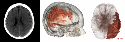
Sample comparison between images used for standard ABC/2 (a.) and 3DI (b. and c.) methods in quantification of subdural hematoma. Figure created by Scott Collins, Diagnostic Imaging.
3D Imaging Research Lab, Department of Diagnostic Imaging, Rhode Island Hospital
The RI Hospital Imaging Research and Education Service (HI-RES) and 3D Lab is a state-of-the-art, 1800 square foot, dry lab facility dedicated to medical image analysis and procedure guidance. The lab supports current research in neurological emergencies, such as cerebroventricular segmentation and 3D segmentation of brain hemorrhage related to TBI and stroke. The lab provides multiple high-end clinical and research workstations, it maintains a conference space with two 80” smart displays, and a computational research simulation and visualization bench within the hospital. The bench space is equipped with hardware and cameras for motion tracking, 3D capture, and stereo display. Imaging research workstations are set up with a variety of scientific software, productivity, design, and programing tools suitable for the AVA project such as Osirix, COMSOL Multiphysics, Matlab, SAS, SPSS, AutoCAD, ITK, VTK, ImageJ, Python, VisualStudio, Xcode, Office, and the Adobe Creative Suite. The lab provides primary workspace for 6 faculty, staff, and graduate student researchers.
Mission
To facilitate interdepartmental collaboration, education, research, and the advancement of clinical care in neurological emergencies.